This Week in Radiology — Oct 24, 2026
Generated Oct 24, 2026 · 11:02
The week's practice-changing Radiology research, summarized for clinicians.
If the audio fails to play, refresh the page to renew the link.
Get next week’s Radiology briefing — free.
In your podcast app, or readable in your inbox with the audio one tap away.
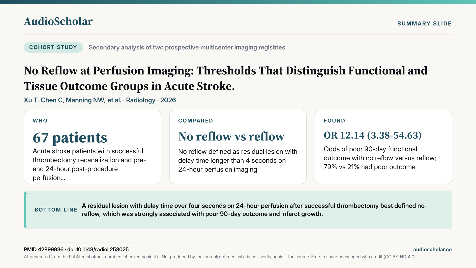
No Reflow at Perfusion Imaging: Thresholds That Distinguish Functional and Tissue Outcome Groups in Acute Stroke.
A residual lesion with delay time over four seconds on 24-hour perfusion after successful thrombectomy best defined no-reflow, which was strongly associated with poor 90-day outcome and infarct growth.
Radiology · 2026 · PubMed

This week’s papers
- 01
Advanced CT Myelography for Spontaneous Cerebrospinal Fluid Leaks.
Localizing spontaneous spinal CSF leaks depends on tailoring CT myelography positioning and technique to the leak type suggested by spine MRI, while recognizing artifacts that mimic leaks.
Carlton Jones L et al. · RadioGraphics · 2026
From the briefingIn RadioGraphics, Carlton Jones and colleagues offer a technical review of advanced CT myelography for spontaneous spinal leaks [1]. Their central argument is that the study has to be tailored to the leak type suspected on spine MRI: ventral and lateral dural tears and CSF venous fistulas each call for different positioning and acquisition strategies, and the review catalogues typical and atypical appearances along with artifacts that can mimic a leak.
Written by AI from the published abstract, for healthcare professionals. Not medical advice; check the paper before acting on it.
- 02
Early Acquisition Timing and CSF Venous Fistula Conspicuity on Lateral Decubitus CT Myelography.
CSF venous fistulas were far more conspicuous at one minute than two minutes after intrathecal injection, though later images added value in some patients, supporting multiphasic imaging.
Ryu B et al. · American Journal of Neuroradiology · 2026
From the briefingA paper by Ryu and colleagues in the American Journal of Neuroradiology addresses one specific variable, acquisition timing on lateral decubitus CT myelography [2]. In 35 patients with high-probability or definite fistulas, three blinded neurointerventional radiologists compared images acquired one minute and two minutes after the start of intrathecal injection. Fistulas met the visibility threshold in close to nine in ten ratings at one minute, against fewer than half of ratings at two minutes, and readers preferred the earlier phase in roughly three quarters of patients. That advantage persisted even after accounting for intrathecal contrast density. Still, the later phase added information in a minority of patients, and the authors are careful to say their data support multiphasic imaging rather than a single universal delay.
Written by AI from the published abstract, for healthcare professionals. Not medical advice; check the paper before acting on it.
- 03
Salvage CT Myelography: Same-Session Repositioning to Localize Spinal CSF Leaks.
In eleven patients, same-session repositioning with extra contrast boluses identified alternative CSF leak types with reasonable procedure times, potentially avoiding repeat myelograms, though evidence remains preliminary.
Mamlouk MD et al. · American Journal of Neuroradiology · 2026
From the briefingAlso in the American Journal of Neuroradiology, Mamlouk and colleagues describe what they call salvage CT myelography [3]: when the suspected leak type isn't found, the patient is repositioned with the needle still in place and given additional small contrast boluses to look for an alternative leak. Across eleven patients at three institutions, the approach identified alternative leaks spanning dural tears and venous fistulas, with a mean procedure time just under an hour and a mean contrast dose of about 12 and a half cc. This is a small technical series without a comparison group, so it shows feasibility rather than a proven reduction in repeat lumbar punctures, but taken together these three papers point toward a more adaptive, multiphase approach to leak localization.
Written by AI from the published abstract, for healthcare professionals. Not medical advice; check the paper before acting on it.
- 04
ASFNR Clinical State of Practice Statement on Imaging of Chronic Subdural Hematoma.
Expert consensus positions noncontrast CT as primary for chronic subdural hematoma, standardizes size and Nakaguchi phenotype reporting, and states phenotype is not yet validated for treatment selection.
Ahmed S et al. · American Journal of Neuroradiology · 2026
From the briefingWith middle meningeal artery embolization now established, the American Society of Functional Neuroradiology has issued a State of Practice statement on imaging of chronic subdural hematoma, published in the American Journal of Neuroradiology and led by Ahmed [4]. The committee positions noncontrast CT as the primary modality, recommends reporting maximum thickness perpendicular to the inner table, midline shift and, where appropriate, volume, and endorses the Nakaguchi framework of homogeneous, laminar, separated and trabecular phenotypes. It is explicit that imaging phenotype has not been validated for treatment selection, and that dual-energy, photon-counting, MRI and arterial spin labeling techniques remain problem-solving tools resting on largely single-center evidence.
Written by AI from the published abstract, for healthcare professionals. Not medical advice; check the paper before acting on it.
- 05
Bilateral Versus Unilateral Middle Meningeal Artery Embolization for Standalone Treatment of Unilateral Chronic Subdural Hematoma.
Bilateral embolization for unilateral subdural hematoma was associated with no index-admission surgical rescues versus about ten percent with unilateral, but overall retreatment did not differ.
Gomez-Paz S et al. · American Journal of Neuroradiology · 2026
From the briefingA multicenter cohort from Gomez-Paz and colleagues, also in the American Journal of Neuroradiology, takes up a procedural question: whether embolizing both middle meningeal arteries helps when the hematoma is on one side [5]. Among 600 patients at 16 centers, surgical rescue during the index admission occurred in about one in ten unilateral cases and in none of the 54 bilateral cases, a difference that held after propensity matching. But overall retreatment did not differ, bilateral cases started with less severe hematomas, and follow-up after discharge was incomplete. The authors themselves conclude the data do not support routine bilateral embolization, and frame contralateral dural supply as a candidate selection marker for prospective trials.
Written by AI from the published abstract, for healthcare professionals. Not medical advice; check the paper before acting on it.
- 06
No Reflow at Perfusion Imaging: Thresholds That Distinguish Functional and Tissue Outcome Groups in Acute Stroke.
A residual lesion with delay time over four seconds on 24-hour perfusion after successful thrombectomy best defined no-reflow, which was strongly associated with poor 90-day outcome and infarct growth.
Xu T et al. · Radiology · 2026
From the briefingIn Radiology, Xu and colleagues tackle the no-reflow phenomenon after successful thrombectomy [6]. Using two prospective multicenter registries, they tested candidate definitions across eight perfusion maps at 24 hours, and found that a residual lesion with delay time longer than four seconds best discriminated poor 90-day outcome. About a fifth of patients met that definition, and among them roughly four in five had a poor outcome, compared with about one in five patients with reflow, along with substantially more infarct growth. The finding replicated in an independent cohort of 54 patients, but both cohorts are small, and this offers an operational definition for research rather than a basis for treatment decisions.
If you only have time for one paper this week, make it the no-reflow study by Xu and colleagues in Radiology [6]. It gives the field a concrete, replicated perfusion definition for a phenomenon that may explain why technically successful thrombectomy so often fails clinically, opening the door to trials targeting the microcirculation.
Written by AI from the published abstract, for healthcare professionals. Not medical advice; check the paper before acting on it.
- 07
Sequential transarterial radioembolization and atezolizumab plus bevacizumab improves survival in advanced hepatocellular carcinoma.
In a retrospective single-center cohort, radioembolization before atezolizumab plus bevacizumab was associated with longer survival than systemic therapy alone, especially with preserved liver function.
Ko Y et al. · European Radiology · 2026
From the briefingIn European Radiology, Ko and colleagues report a single-center retrospective comparison of yttrium-90 radioembolization followed by atezolizumab plus bevacizumab versus the drug combination alone in 156 systemic therapy-naive patients with advanced hepatocellular carcinoma [7]. Median overall survival was roughly 14 months with the sequential approach against about six months with systemic therapy alone, and the association held after propensity matching and weighting. Progression-free survival was similar overall, but both survival measures favored the combined strategy in patients with well-preserved liver function. Selection bias is the obvious concern in a retrospective series like this, so the findings generate a hypothesis for trials rather than settle the question.
Written by AI from the published abstract, for healthcare professionals. Not medical advice; check the paper before acting on it.
- 08
CT-based Node-RADS for standardized evaluation of lymph node metastasis in rectal cancer and its clinicopathological correlates.
CT-based Node-RADS showed good discrimination and substantial inter-reader agreement for nodal metastasis in 459 treatment-naive rectal cancer patients, with scores correlating with T stage and lymphovascular invasion.
Xiao X et al. · European Journal of Radiology · 2026
From the briefingIn the European Journal of Radiology, Xiao and colleagues evaluated CT-based Node-RADS in 459 treatment-naive rectal cancer patients with surgical pathology [8]. A score of three or more captured close to nine in ten node-positive patients at the cost of modest specificity, while a threshold of four balanced sensitivity and specificity in the mid seventies. Overall discrimination was good, inter-reader agreement was substantial, and scores tracked with T stage, N stage and lymphovascular invasion. It is a single-center study, and MRI remains the mainstay for local rectal staging, but it supports Node-RADS as a reproducible reporting framework on CT.
Written by AI from the published abstract, for healthcare professionals. Not medical advice; check the paper before acting on it.
- 09
Timeliness of an Expedited Diagnostic Pathway for Screening Mammography Recalls.
Radiologist-flagged expedited mammography recalls reached diagnostic imaging in seven rather than 25 days and had very high malignancy yield, supporting prioritization of diagnostic resources.
Panigrahi B et al. · Journal of the American College of Radiology · 2026
From the briefingOn the breast side, Panigrahi and colleagues in the Journal of the American College of Radiology describe an expedited recall pathway, built in the electronic record, that let radiologists flag highly suspicious BI-RADS 0 screening findings for reserved appointments within a week [9]. Only about one percent of recalls were expedited, but those patients reached diagnostic imaging in a median of seven days rather than 25, and screening-to-biopsy time fell by about three weeks. Radiologists picked well: roughly nine in ten expedited recalls that went to biopsy recommendation proved malignant, versus about one in twenty in the standard pathway. The study does not show any effect on stage or outcomes, but it suggests radiologist triage at screening is accurate enough to justify prioritizing diagnostic slots.
Written by AI from the published abstract, for healthcare professionals. Not medical advice; check the paper before acting on it.
- 10
Intraindividual Comparison of Radiation Dose and Image Quality: Photon-counting versus Energy-integrating Detector CT in Obesity.
In patients with obesity scanned on both systems, photon-counting CT delivered higher signal-to-noise and better reader-rated image quality at lower radiation dose than conventional detector CT.
Hegmanns J et al. · Radiology · 2026
From the briefingFinally, a technology paper in Radiology connects back to the photon-counting CT mentioned in the subdural guidance. Hegmanns and colleagues compared photon-counting and conventional energy-integrating detector CT within the same 51 patients with obesity [10]. Photon-counting delivered higher signal-to-noise in vessels and most organs, better reader-rated quality with fewer artifacts, and did so at roughly 30 percent lower dose. The image quality advantage grew with body mass index. The design is retrospective and modest in size, but the intraindividual comparison makes this a fairly clean signal in a population where dose and image quality typically trade off.
Written by AI from the published abstract, for healthcare professionals. Not medical advice; check the paper before acting on it.
The full briefing
This AudioScholar briefing is generated by artificial intelligence for healthcare professionals and trainees. It is not medical advice.
Welcome to This Week in Radiology. This week we're covering 10 notable papers spanning spinal fluid leak imaging, neurovascular imaging and intervention, and oncologic and breast imaging workflows, with a look at photon-counting CT along the way. Let's dive in.
We start with spontaneous intracranial hypotension, where three papers this week focus on how CT myelography is actually performed. In RadioGraphics, Carlton Jones and colleagues offer a technical review of advanced CT myelography for spontaneous spinal leaks [1]. Their central argument is that the study has to be tailored to the leak type suspected on spine MRI: ventral and lateral dural tears and CSF venous fistulas each call for different positioning and acquisition strategies, and the review catalogues typical and atypical appearances along with artifacts that can mimic a leak. A paper by Ryu and colleagues in the American Journal of Neuroradiology addresses one specific variable, acquisition timing on lateral decubitus CT myelography [2]. In 35 patients with high-probability or definite fistulas, three blinded neurointerventional radiologists compared images acquired one minute and two minutes after the start of intrathecal injection. Fistulas met the visibility threshold in close to nine in ten ratings at one minute, against fewer than half of ratings at two minutes, and readers preferred the earlier phase in roughly three quarters of patients. That advantage persisted even after accounting for intrathecal contrast density. Still, the later phase added information in a minority of patients, and the authors are careful to say their data support multiphasic imaging rather than a single universal delay. Also in the American Journal of Neuroradiology, Mamlouk and colleagues describe what they call salvage CT myelography [3]: when the suspected leak type isn't found, the patient is repositioned with the needle still in place and given additional small contrast boluses to look for an alternative leak. Across eleven patients at three institutions, the approach identified alternative leaks spanning dural tears and venous fistulas, with a mean procedure time just under an hour and a mean contrast dose of about 12 and a half cc. This is a small technical series without a comparison group, so it shows feasibility rather than a proven reduction in repeat lumbar punctures, but taken together these three papers point toward a more adaptive, multiphase approach to leak localization.
Our second theme moves inside the skull, to chronic subdural hematoma and the aftermath of stroke thrombectomy. With middle meningeal artery embolization now established, the American Society of Functional Neuroradiology has issued a State of Practice statement on imaging of chronic subdural hematoma, published in the American Journal of Neuroradiology and led by Ahmed [4]. The committee positions noncontrast CT as the primary modality, recommends reporting maximum thickness perpendicular to the inner table, midline shift and, where appropriate, volume, and endorses the Nakaguchi framework of homogeneous, laminar, separated and trabecular phenotypes. It is explicit that imaging phenotype has not been validated for treatment selection, and that dual-energy, photon-counting, MRI and arterial spin labeling techniques remain problem-solving tools resting on largely single-center evidence. A multicenter cohort from Gomez-Paz and colleagues, also in the American Journal of Neuroradiology, takes up a procedural question: whether embolizing both middle meningeal arteries helps when the hematoma is on one side [5]. Among 600 patients at 16 centers, surgical rescue during the index admission occurred in about one in ten unilateral cases and in none of the 54 bilateral cases, a difference that held after propensity matching. But overall retreatment did not differ, bilateral cases started with less severe hematomas, and follow-up after discharge was incomplete. The authors themselves conclude the data do not support routine bilateral embolization, and frame contralateral dural supply as a candidate selection marker for prospective trials. In Radiology, Xu and colleagues tackle the no-reflow phenomenon after successful thrombectomy [6]. Using two prospective multicenter registries, they tested candidate definitions across eight perfusion maps at 24 hours, and found that a residual lesion with delay time longer than four seconds best discriminated poor 90-day outcome. About a fifth of patients met that definition, and among them roughly four in five had a poor outcome, compared with about one in five patients with reflow, along with substantially more infarct growth. The finding replicated in an independent cohort of 54 patients, but both cohorts are small, and this offers an operational definition for research rather than a basis for treatment decisions.
Our third theme covers oncologic imaging and breast screening, where standardization and pathway design took center stage. In European Radiology, Ko and colleagues report a single-center retrospective comparison of yttrium-90 radioembolization followed by atezolizumab plus bevacizumab versus the drug combination alone in 156 systemic therapy-naive patients with advanced hepatocellular carcinoma [7]. Median overall survival was roughly 14 months with the sequential approach against about six months with systemic therapy alone, and the association held after propensity matching and weighting. Progression-free survival was similar overall, but both survival measures favored the combined strategy in patients with well-preserved liver function. Selection bias is the obvious concern in a retrospective series like this, so the findings generate a hypothesis for trials rather than settle the question. In the European Journal of Radiology, Xiao and colleagues evaluated CT-based Node-RADS in 459 treatment-naive rectal cancer patients with surgical pathology [8]. A score of three or more captured close to nine in ten node-positive patients at the cost of modest specificity, while a threshold of four balanced sensitivity and specificity in the mid seventies. Overall discrimination was good, inter-reader agreement was substantial, and scores tracked with T stage, N stage and lymphovascular invasion. It is a single-center study, and MRI remains the mainstay for local rectal staging, but it supports Node-RADS as a reproducible reporting framework on CT. On the breast side, Panigrahi and colleagues in the Journal of the American College of Radiology describe an expedited recall pathway, built in the electronic record, that let radiologists flag highly suspicious BI-RADS 0 screening findings for reserved appointments within a week [9]. Only about one percent of recalls were expedited, but those patients reached diagnostic imaging in a median of seven days rather than 25, and screening-to-biopsy time fell by about three weeks. Radiologists picked well: roughly nine in ten expedited recalls that went to biopsy recommendation proved malignant, versus about one in twenty in the standard pathway. The study does not show any effect on stage or outcomes, but it suggests radiologist triage at screening is accurate enough to justify prioritizing diagnostic slots.
Finally, a technology paper in Radiology connects back to the photon-counting CT mentioned in the subdural guidance. Hegmanns and colleagues compared photon-counting and conventional energy-integrating detector CT within the same 51 patients with obesity [10]. Photon-counting delivered higher signal-to-noise in vessels and most organs, better reader-rated quality with fewer artifacts, and did so at roughly 30 percent lower dose. The image quality advantage grew with body mass index. The design is retrospective and modest in size, but the intraindividual comparison makes this a fairly clean signal in a population where dose and image quality typically trade off.
If you only have time for one paper this week, make it the no-reflow study by Xu and colleagues in Radiology [6]. It gives the field a concrete, replicated perfusion definition for a phenomenon that may explain why technically successful thrombectomy so often fails clinically, opening the door to trials targeting the microcirculation.
Here is what this week's evidence adds up to in Radiology. First, CT myelography for spinal leaks is becoming more tailored and adaptive: early acquisition improves fistula conspicuity and same-session repositioning appears feasible, though both rest on small retrospective series and neither establishes a single optimal protocol. Second, imaging of chronic subdural hematoma now has consensus reporting standards, but phenotype is not yet validated for selecting patients, and the signal favoring bilateral embolization is observational and not supported by the authors for routine use. Third, a delay time over four seconds on 24-hour perfusion is emerging as a reproducible marker of no-reflow after thrombectomy, strongly linked to poor outcome, but it remains a research definition. Fourth, in oncology, radioembolization before atezolizumab and bevacizumab was associated with longer survival in patients with preserved liver function, a retrospective finding that awaits randomized confirmation, while CT Node-RADS shows reproducible performance in rectal cancer. And fifth, radiologist-flagged expedited recalls and photon-counting CT in obesity both show measurable gains in efficiency and quality, though neither has yet been linked to patient outcomes.
That's your roundup for This Week in Radiology. The full transcript and references are available on the episode page. This is an AI-curated summary — for clinical decisions, always consult primary sources and current guidelines. See you next week.
If this weekly briefing is useful, follow the show in your podcast app so new episodes arrive automatically. And think of one colleague — in any specialty — who never has time to keep up with the literature. Tell them about AudioScholar: a free ten-minute weekly for every specialty, to listen to in any podcast app, or to read at audioscholar dot C C.
This is an automated summary generated by artificial intelligence, which can make mistakes. Always review the original source materials.
Spot something worth flagging?
Get this every week in your podcast app — free.
New radiology episodes land in your feed automatically — listen on your commute.