This Week in Ophthalmology — Oct 25, 2026
Generated Oct 25, 2026 · 11:32
The week's practice-changing Ophthalmology research, summarized for clinicians.
If the audio fails to play, refresh the page to renew the link.
Get next week’s Ophthalmology briefing — free.
In your podcast app, or readable in your inbox with the audio one tap away.
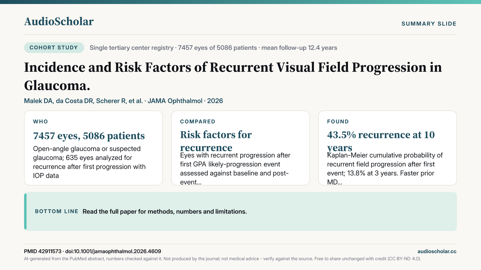
Incidence and Risk Factors of Recurrent Visual Field Progression in Glaucoma.
After a first confirmed visual field progression, about 44 percent of glaucomatous eyes recurred by ten years, with faster prior loss, higher post-event pressure, and older age raising risk.
JAMA Ophthalmology · 2026 · PubMed

This week’s papers
- 01
Incidence and Risk Factors of Recurrent Visual Field Progression in Glaucoma.
After a first confirmed visual field progression, about 44 percent of glaucomatous eyes recurred by ten years, with faster prior loss, higher post-event pressure, and older age raising risk.
Malek DA et al. · JAMA Ophthalmology · 2026
From the briefingIn JAMA Ophthalmology, Malek and colleagues drew on the Bascom Palmer registry, following more than seven thousand eyes for an average of about twelve years, to examine what happens after a first confirmed visual field progression event on Guided Progression Analysis [1]. Among eyes with adequate follow-up after that first event, recurrence was common: roughly one in seven eyes had progressed again by three years, and the cumulative estimate climbed to just over forty percent by ten years. Four features independently marked the higher-risk eyes. Faster mean deviation loss before the first event raised the hazard by about seventy percent for each additional decibel per year, and higher average pressure after the event, less advanced damage at first progression, and older age all added risk. Pressure lowering mattered in the expected direction, with each additional ten percent reduction in pressure linked to roughly an eleven percent lower hazard. Put simply, eyes that were losing more than one decibel a year before the first event recurred at nearly twice the rate of slower eyes, about 43 versus 23 percent. This is single-centre, observational data, but it gives quantitative support to the idea that the pre-event rate of loss, not just the event itself, identifies who may warrant closer monitoring and more aggressive pressure targets.
If you only have time for one paper this week, make it the recurrent visual field progression study from JAMA Ophthalmology [1]. It addresses a question that arises in almost every glaucoma clinic, what happens after the first confirmed progression, and gives long-term, quantitative evidence on which eyes are most likely to progress again.
Written by AI from the published abstract, for healthcare professionals. Not medical advice; check the paper before acting on it.
- 02
Association Between Systemic TNF-α Inhibitor Use and Risk of Primary Open-Angle Glaucoma.
In matched health-record data, systemic TNF-alpha inhibitor users had about 30 percent lower ten-year risk of open-angle glaucoma diagnosis, an observational signal implicating inflammation in neurodegeneration.
Hong AT et al. · American Journal of Ophthalmology · 2026
From the briefingAlongside it, in the American Journal of Ophthalmology, Hong and colleagues used electronic health records from 70 health care organizations in the United States to compare nearly twelve thousand propensity-matched pairs of patients with and without systemic TNF-alpha inhibitor exposure [2]. Over ten years, new primary open-angle glaucoma diagnoses were less frequent among inhibitor users, 2.0 versus 3.3 percent, a reduction of about thirty percent in hazard, with adalimumab and infliximab each showing a signal and no significant differences between agents. The authors frame this as support for inflammation in glaucomatous neurodegeneration, but the diagnoses rest on billing codes and residual confounding is likely, so this is hypothesis-generating rather than a reason to view these drugs as glaucoma therapy.
Written by AI from the published abstract, for healthcare professionals. Not medical advice; check the paper before acting on it.
- 03
Longitudinal analysis of structural biomarkers for disease progression in intermediate age-related macular degeneration: a MACUSTAR study report.
In 577 eyes with intermediate macular degeneration, central drusenoid deposits, detachments, non-exudative neovascularization and atrophy predicted progression, with distinct biomarkers and topography separating atrophic from neovascular conversion.
Goerdt L et al. · Ophthalmology Retina · 2026
From the briefingIn Ophthalmology Retina, Goerdt and colleagues report the longitudinal arm of the European multicentre MACUSTAR study, following 577 eyes with intermediate disease for up to four years with standardized multimodal imaging and deep-learning quantification of OCT features [3]. Eighty-two eyes progressed, 37 to geographic atrophy and 45 to neovascular disease. Within the central three millimetres, subretinal drusenoid deposits, pigment epithelial detachment, non-exudative neovascularization and complete outer retinal atrophy conferred overall risk, while pigmentary abnormalities, drusen complex volume and incomplete or complete atrophy pointed specifically toward geographic atrophy. Only the volume of non-exudative neovascularization signalled conversion to neovascular disease. The key nuance is location: the same biomarker carried different prognostic weight depending on where in the macula it sat, which the authors argue should shape clinical trial design.
Written by AI from the published abstract, for healthcare professionals. Not medical advice; check the paper before acting on it.
- 04
Risk Factors for Retinal Pigment Epithelium Tears in Patients with Neovascular Age-Related Macular Degeneration: A Systematic Review and Meta-Analysis.
Across 16 studies, taller and larger pigment epithelial detachments were most consistently associated with RPE tears in neovascular macular degeneration, though overall certainty of evidence was very low.
Al-Burak SA et al. · American Journal of Ophthalmology · 2026
From the briefingOnce neovascular disease is established, a different structural question arises, and Al-Burak and colleagues addressed it in the American Journal of Ophthalmology with a meta-analysis of 16 studies and about four thousand eyes on risk factors for retinal pigment epithelium tears [4]. Eyes that went on to tear had baseline pigment epithelial detachments roughly 200 microns taller and several square millimetres larger, and fibrovascular detachments, sub-RPE clefts, subretinal fluid and occult or haemorrhagic neovascularization were also associated. The choice of anti-VEGF agent was not consistently linked to tears. The authors are candid that certainty is very low, the studies largely retrospective and unadjusted, so detachment height is best read as a consistent association that may inform counselling, not a validated predictor.
Written by AI from the published abstract, for healthcare professionals. Not medical advice; check the paper before acting on it.
- 05
OCT to Assess Risk of Progression of Multiple Sclerosis: A Report by the American Academy of Ophthalmology.
Thinner or faster-thinning retinal nerve fibre and ganglion cell layers on OCT were associated with future multiple sclerosis disability, relapse and MRI activity, supporting biomarker development.
Moss HE et al. · Ophthalmology · 2026
From the briefingIn Ophthalmology, a report from the American Academy of Ophthalmology led by Moss reviewed 21 studies on whether OCT predicts multiple sclerosis progression [5]. Only two of those studies reached level one evidence, but across the body of work, thinner peripapillary nerve fibre layer and ganglion cell layer, faster thinning, and larger differences between the eyes were associated with future disability, relapses and MRI activity, including in radiologically and clinically isolated syndromes, and the association held in multivariable models. The panel concludes the evidence supports developing these measures as biomarkers, while flagging gaps in demographic diversity, cross-device comparability, and how OCT should be weighed against MRI.
Written by AI from the published abstract, for healthcare professionals. Not medical advice; check the paper before acting on it.
- 06
Systematic Review of Clinical Utility of Multimodal Imaging in Infectious Posterior Uveitis. Multimodal imaging in Uveitis (MUV) Taskforce Report 12.
Across 122 studies of five infectious posterior uveitides, fundus photography and OCT achieved high certainty for most clinical uses, while acute retinal necrosis imaging evidence remained comparatively limited.
Singh Parmar UP et al. · Ophthalmology Retina · 2026
From the briefingAlso in Ophthalmology Retina, the Multimodal Imaging in Uveitis taskforce, in a report led by Singh Parmar, systematically reviewed 122 studies covering toxoplasmosis, cytomegalovirus retinitis, tuberculosis, syphilis and acute retinal necrosis [6]. Colour fundus photography and OCT achieved high certainty across most domains in four of the five entities, autofluorescence was strongest for disease activity, fluorescein angiography for complications, while OCT angiography and indocyanine green angiography had thinner support; acute retinal necrosis remains the least well studied.
Written by AI from the published abstract, for healthcare professionals. Not medical advice; check the paper before acting on it.
- 07
Artificial intelligence in diabetic retinopathy: From deep learning to foundation models, clinical translation, and real-world deployment.
Real-world success of AI diabetic retinopathy screening depends on image gradability, thresholds, external validation, pigmentation-related disparities and referral capacity, not benchmark accuracy alone, according to this narrative review.
Haobijam B et al. · Survey of Ophthalmology · 2026
From the briefingRounding out the theme, a structured narrative review in Survey of Ophthalmology by Haobijam and colleagues traces artificial intelligence for diabetic retinopathy from convolutional networks to retina-specific foundation models [7]. Its central argument is that real-world screening success depends less on benchmark accuracy than on image gradability, appropriate thresholds, external validation across devices and populations, performance disparities related to fundus pigmentation, and referral capacity. It is a synthesis rather than new data, but a useful map of where deployment evidence is solid and where it is not.
Written by AI from the published abstract, for healthcare professionals. Not medical advice; check the paper before acting on it.
- 08
Efficacy of Oral Belzutifan for Retinal Capillary Hemangioblastomas in von Hippel-Lindau Disease.
Oral belzutifan shrank active retinal hemangioblastoma area by about three quarters at one year in von Hippel-Lindau disease, but over 70 percent of eyes relapsed after stopping.
Ji MH et al. · Ophthalmology Retina · 2026
From the briefingIn Ophthalmology Retina, Ji and colleagues at the National Eye Institute describe 33 patients with von Hippel-Lindau disease, 42 eyes, treated with oral belzutifan [8]. Total active retinal hemangioblastoma area fell by about three quarters at one year and about eighty percent at two years, and no new tumours appeared on treatment. The sobering counterpoint is that more than seventy percent of eyes showed relapse after the drug was stopped. The authors see a role for juxtapapillary tumours and as adjunctive therapy for large or multifocal lesions, with ongoing therapy likely needed; it remains a retrospective, single-centre series without a comparator.
Written by AI from the published abstract, for healthcare professionals. Not medical advice; check the paper before acting on it.
- 09
Supervised Reduction Protocol for Management of Herpetic Stromal Keratitis.
Under closely supervised steroid tapering for herpetic stromal keratitis, most recurrences followed steroid discontinuation and were often asymptomatic at detection, though superiority over other approaches is unproven.
Shetty S et al. · Cornea · 2026
From the briefingIn Cornea, Shetty and colleagues describe a supervised reduction protocol for herpetic stromal keratitis in 53 patients, tapering steroids and antivirals only under direct slit-lamp supervision [9]. Recurrence occurred in a third of patients with herpes simplex disease and half of patients with zoster disease, most recurrences followed steroid discontinuation, none occurred on four-times-daily dosing, and most recurrences were asymptomatic when detected, with vision stable through to quiescence. The authors explicitly note this single-physician series cannot show superiority over other approaches.
Written by AI from the published abstract, for healthcare professionals. Not medical advice; check the paper before acting on it.
- 10
Morgagnian Cataract: Clinical Features, Surgical Management, and Outcomes.
Morgagnian cataracts often presented with hand-motion vision, raised pressure and zonular weakness; surgery carried a one-in-five capsule rupture rate, yet half of operated eyes reached 20/40.
Kang S et al. · American Journal of Ophthalmology · 2026
From the briefingFinally, in the American Journal of Ophthalmology, Kang and colleagues reviewed 50 eyes with Morgagnian cataract [10]. Most eyes presented at hand motions or worse, close to half of eyes had lens-induced pressure elevation, and zonular weakness was frequent. Among operated eyes, posterior capsule rupture occurred in about one in five and only four in ten received an in-the-bag lens, yet half of the operated eyes reached 20/40 or better and pressure fell substantially, though half of those eyes still needed glaucoma drops. The data quantify surgical risk for counselling, within the limits of a modest retrospective tertiary series.
Written by AI from the published abstract, for healthcare professionals. Not medical advice; check the paper before acting on it.
The full briefing
This AudioScholar briefing is generated by artificial intelligence for healthcare professionals and trainees. It is not medical advice.
Welcome to This Week in Ophthalmology. This week we're covering 10 notable papers spanning glaucoma risk stratification, imaging biomarkers that forecast disease in the retina and beyond, and treatment of difficult anterior and posterior segment conditions. Let's dive in.
We start with glaucoma, where two large cohort studies ask who keeps losing vision and what might be quietly protecting others. In JAMA Ophthalmology, Malek and colleagues drew on the Bascom Palmer registry, following more than seven thousand eyes for an average of about twelve years, to examine what happens after a first confirmed visual field progression event on Guided Progression Analysis [1]. Among eyes with adequate follow-up after that first event, recurrence was common: roughly one in seven eyes had progressed again by three years, and the cumulative estimate climbed to just over forty percent by ten years. Four features independently marked the higher-risk eyes. Faster mean deviation loss before the first event raised the hazard by about seventy percent for each additional decibel per year, and higher average pressure after the event, less advanced damage at first progression, and older age all added risk. Pressure lowering mattered in the expected direction, with each additional ten percent reduction in pressure linked to roughly an eleven percent lower hazard. Put simply, eyes that were losing more than one decibel a year before the first event recurred at nearly twice the rate of slower eyes, about 43 versus 23 percent. This is single-centre, observational data, but it gives quantitative support to the idea that the pre-event rate of loss, not just the event itself, identifies who may warrant closer monitoring and more aggressive pressure targets. Alongside it, in the American Journal of Ophthalmology, Hong and colleagues used electronic health records from 70 health care organizations in the United States to compare nearly twelve thousand propensity-matched pairs of patients with and without systemic TNF-alpha inhibitor exposure [2]. Over ten years, new primary open-angle glaucoma diagnoses were less frequent among inhibitor users, 2.0 versus 3.3 percent, a reduction of about thirty percent in hazard, with adalimumab and infliximab each showing a signal and no significant differences between agents. The authors frame this as support for inflammation in glaucomatous neurodegeneration, but the diagnoses rest on billing codes and residual confounding is likely, so this is hypothesis-generating rather than a reason to view these drugs as glaucoma therapy.
Turning to the macula, two papers refine which structural features on imaging predict trouble in age-related macular degeneration. In Ophthalmology Retina, Goerdt and colleagues report the longitudinal arm of the European multicentre MACUSTAR study, following 577 eyes with intermediate disease for up to four years with standardized multimodal imaging and deep-learning quantification of OCT features [3]. Eighty-two eyes progressed, 37 to geographic atrophy and 45 to neovascular disease. Within the central three millimetres, subretinal drusenoid deposits, pigment epithelial detachment, non-exudative neovascularization and complete outer retinal atrophy conferred overall risk, while pigmentary abnormalities, drusen complex volume and incomplete or complete atrophy pointed specifically toward geographic atrophy. Only the volume of non-exudative neovascularization signalled conversion to neovascular disease. The key nuance is location: the same biomarker carried different prognostic weight depending on where in the macula it sat, which the authors argue should shape clinical trial design. Once neovascular disease is established, a different structural question arises, and Al-Burak and colleagues addressed it in the American Journal of Ophthalmology with a meta-analysis of 16 studies and about four thousand eyes on risk factors for retinal pigment epithelium tears [4]. Eyes that went on to tear had baseline pigment epithelial detachments roughly 200 microns taller and several square millimetres larger, and fibrovascular detachments, sub-RPE clefts, subretinal fluid and occult or haemorrhagic neovascularization were also associated. The choice of anti-VEGF agent was not consistently linked to tears. The authors are candid that certainty is very low, the studies largely retrospective and unadjusted, so detachment height is best read as a consistent association that may inform counselling, not a validated predictor.
Our third theme widens the lens to imaging as a window on systemic and infectious disease, and to the infrastructure needed to use it. In Ophthalmology, a report from the American Academy of Ophthalmology led by Moss reviewed 21 studies on whether OCT predicts multiple sclerosis progression [5]. Only two of those studies reached level one evidence, but across the body of work, thinner peripapillary nerve fibre layer and ganglion cell layer, faster thinning, and larger differences between the eyes were associated with future disability, relapses and MRI activity, including in radiologically and clinically isolated syndromes, and the association held in multivariable models. The panel concludes the evidence supports developing these measures as biomarkers, while flagging gaps in demographic diversity, cross-device comparability, and how OCT should be weighed against MRI. Also in Ophthalmology Retina, the Multimodal Imaging in Uveitis taskforce, in a report led by Singh Parmar, systematically reviewed 122 studies covering toxoplasmosis, cytomegalovirus retinitis, tuberculosis, syphilis and acute retinal necrosis [6]. Colour fundus photography and OCT achieved high certainty across most domains in four of the five entities, autofluorescence was strongest for disease activity, fluorescein angiography for complications, while OCT angiography and indocyanine green angiography had thinner support; acute retinal necrosis remains the least well studied. Rounding out the theme, a structured narrative review in Survey of Ophthalmology by Haobijam and colleagues traces artificial intelligence for diabetic retinopathy from convolutional networks to retina-specific foundation models [7]. Its central argument is that real-world screening success depends less on benchmark accuracy than on image gradability, appropriate thresholds, external validation across devices and populations, performance disparities related to fundus pigmentation, and referral capacity. It is a synthesis rather than new data, but a useful map of where deployment evidence is solid and where it is not.
Our final theme concerns treatment of uncommon but sight-threatening conditions, where evidence inevitably comes from case series. In Ophthalmology Retina, Ji and colleagues at the National Eye Institute describe 33 patients with von Hippel-Lindau disease, 42 eyes, treated with oral belzutifan [8]. Total active retinal hemangioblastoma area fell by about three quarters at one year and about eighty percent at two years, and no new tumours appeared on treatment. The sobering counterpoint is that more than seventy percent of eyes showed relapse after the drug was stopped. The authors see a role for juxtapapillary tumours and as adjunctive therapy for large or multifocal lesions, with ongoing therapy likely needed; it remains a retrospective, single-centre series without a comparator. In Cornea, Shetty and colleagues describe a supervised reduction protocol for herpetic stromal keratitis in 53 patients, tapering steroids and antivirals only under direct slit-lamp supervision [9]. Recurrence occurred in a third of patients with herpes simplex disease and half of patients with zoster disease, most recurrences followed steroid discontinuation, none occurred on four-times-daily dosing, and most recurrences were asymptomatic when detected, with vision stable through to quiescence. The authors explicitly note this single-physician series cannot show superiority over other approaches. Finally, in the American Journal of Ophthalmology, Kang and colleagues reviewed 50 eyes with Morgagnian cataract [10]. Most eyes presented at hand motions or worse, close to half of eyes had lens-induced pressure elevation, and zonular weakness was frequent. Among operated eyes, posterior capsule rupture occurred in about one in five and only four in ten received an in-the-bag lens, yet half of the operated eyes reached 20/40 or better and pressure fell substantially, though half of those eyes still needed glaucoma drops. The data quantify surgical risk for counselling, within the limits of a modest retrospective tertiary series.
If you only have time for one paper this week, make it the recurrent visual field progression study from JAMA Ophthalmology [1]. It addresses a question that arises in almost every glaucoma clinic, what happens after the first confirmed progression, and gives long-term, quantitative evidence on which eyes are most likely to progress again.
Here is what this week's evidence adds up to in Ophthalmology. First, a first glaucoma progression event appears to be the start rather than the end of risk, with a large but single-centre cohort tying recurrence to prior rate of loss, post-event pressure and age; prospective validation of risk-targeted intensification is still lacking. Second, OCT structural biomarkers are maturing as prognostic tools, in intermediate macular degeneration where location matters, and in multiple sclerosis where the Academy now supports biomarker development, though device comparability and integration with MRI remain open. Third, tall, large pigment epithelial detachments are the most consistent association with RPE tears, but certainty is very low. Fourth, belzutifan shows substantial regression of retinal hemangioblastomas in a retrospective series, with frequent relapse off drug raising unresolved questions about duration of therapy. And the apparent glaucoma protection with TNF-alpha inhibitors is an observational signal that needs confirmation before it carries clinical weight.
That's your roundup for This Week in Ophthalmology. The full transcript and references are available on the episode page. This is an AI-curated summary — for clinical decisions, always consult primary sources and current guidelines. See you next week.
If this weekly briefing is useful, follow the show in your podcast app so new episodes arrive automatically. And think of one colleague — in any specialty — who never has time to keep up with the literature. Tell them about AudioScholar: a free ten-minute weekly for every specialty, to listen to in any podcast app, or to read at audioscholar dot C C.
This is an automated summary generated by artificial intelligence, which can make mistakes. Always review the original source materials.
Spot something worth flagging?
Get this every week in your podcast app — free.
New ophthalmology episodes land in your feed automatically — listen on your commute.