This Week in Radiology — Oct 17, 2026
Generated Oct 17, 2026 · 11:49
The week's practice-changing Radiology research, summarized for clinicians.
If the audio fails to play, refresh the page to renew the link.
Get next week’s Radiology briefing — free.
In your podcast app, or readable in your inbox with the audio one tap away.
Artificial Intelligence for Prostate Cancer Detection and Screening on MRI in Global Populations: The SCARLET-1 Study.
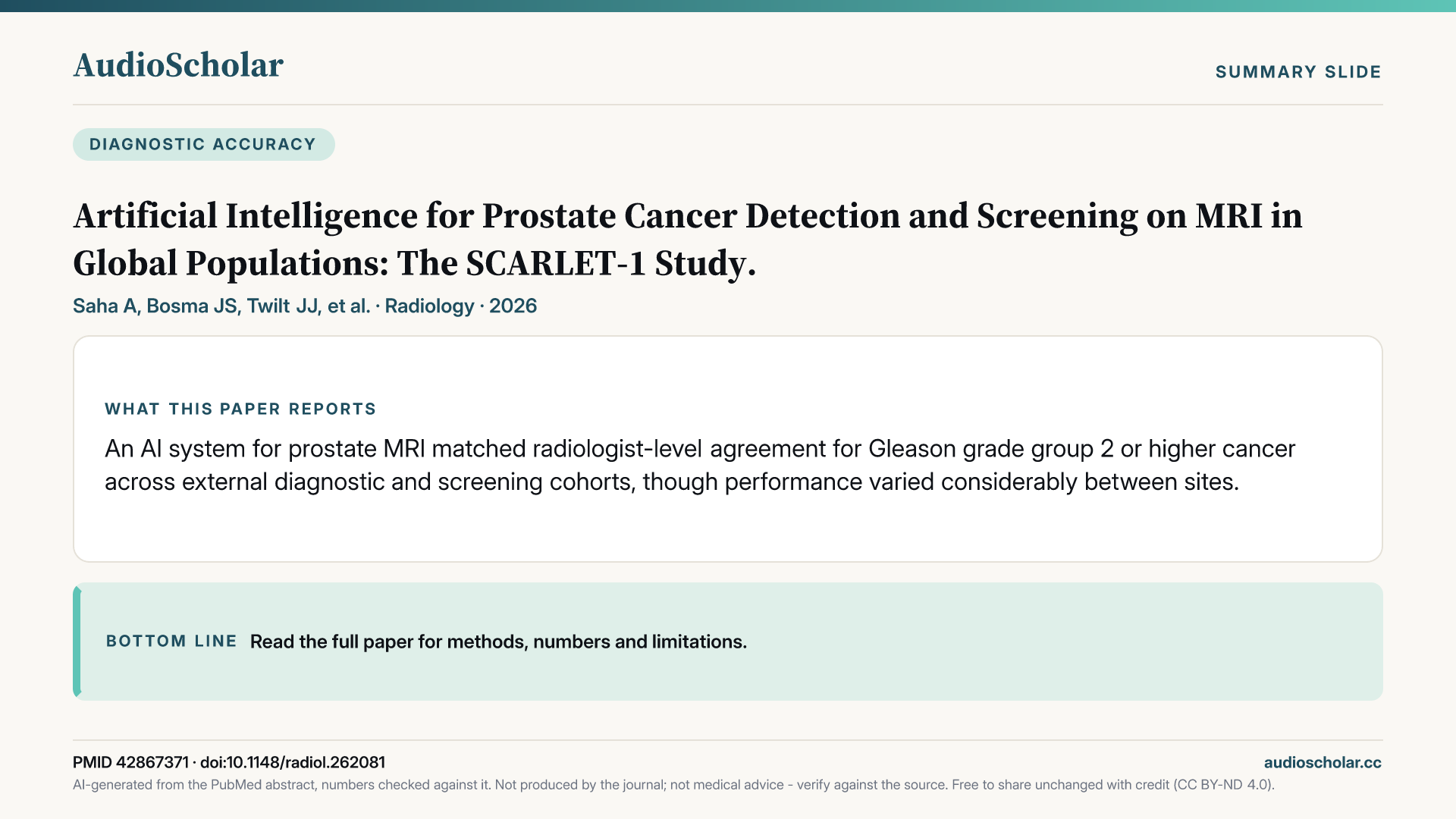
An AI system for prostate MRI matched radiologist-level agreement for Gleason grade group 2 or higher cancer across external diagnostic and screening cohorts, though performance varied considerably between sites.
Radiology · 2026 · PubMed

This week’s papers
- 01
Artificial Intelligence for Prostate Cancer Detection and Screening on MRI in Global Populations: The SCARLET-1 Study.
An AI system for prostate MRI matched radiologist-level agreement for Gleason grade group 2 or higher cancer across external diagnostic and screening cohorts, though performance varied considerably between sites.
Saha A et al. · Radiology · 2026
From the briefingIn Radiology, Saha and colleagues report the SCARLET-1 study, a large retrospective validation of an artificial intelligence system for detecting Gleason grade group 2 or higher prostate cancer [1]. The system was trained and internally tested on more than twenty thousand MRI examinations from 14 countries, then tested externally on close to two thousand patients from 20 further cities, including both a diagnostic cohort and two organized screening cohorts. Agreement between the algorithm and standard-of-care diagnoses ran from about 83 to about 88 percent across the three external datasets, meeting the prespecified criteria for interchangeability with an average radiologist in every one. Discrimination was strong, with areas under the curve in the mid to high eighties. Performance held steady across age, risk group, tumour location and ethnicity, but the variation between sites was substantial: just over a quarter of the 25 sites fell below an area under the curve of 0.80, and lower image quality was associated with weaker performance. That site and quality dependence is arguably the most practical finding, because it suggests local validation and image quality control matter before any deployment. The design is retrospective, so the evidence speaks to diagnostic agreement rather than patient outcomes.
If you only have time for one paper this week, make it the SCARLET-1 study in Radiology [1]. It is the broadest external test yet of prostate MRI artificial intelligence in true screening populations, and its site-to-site variation reopens the question of how such tools should be validated locally before they carry real reading load.
Written by AI from the published abstract, for healthcare professionals. Not medical advice; check the paper before acting on it.
- 02
Prostate cancer screening with MRI: insights from the ESUR Prostate MRI Working Group.
European experts conclude MRI-based prostate screening, especially biparametric MRI after PSA, improves significant cancer detection and reduces unnecessary biopsies, while long-term mortality evidence remains limited.
Brembilla G et al. · European Radiology · 2026
From the briefingFraming all this, a special report in European Radiology from the ESUR Prostate MRI Working Group, led by Brembilla, reviews MRI in prostate screening [2]. The group concludes that MRI-based strategies appear to improve detection of clinically significant cancer while reducing unnecessary biopsies and overdiagnosis, that biparametric MRI is best suited to screening because of its speed and lower cost, and that the strongest evidence supports MRI as a reflex test after a PSA result rather than an MRI-first approach. They are candid that mortality data and interval cancer rates remain limited, and they name capacity, cost and quality assurance as the main barriers, with artificial intelligence flagged as one possible efficiency gain, which is exactly the gap SCARLET-1 tries to address.
Written by AI from the published abstract, for healthcare professionals. Not medical advice; check the paper before acting on it.
- 03
Supplemental Breast Cancer Screening with Contrast-enhanced Mammography or Abbreviated MRI for Extremely Dense Breasts: The DENSE-2 Trial.
The randomized DENSE-2 trial, embedded in Dutch screening, will test whether contrast-enhanced mammography or abbreviated MRI reduces interval cancers in women with extremely dense breasts; results are pending.
van Grinsven SEL et al. · Radiology · 2026
From the briefingIn Radiology, van Grinsven and colleagues describe the design of the DENSE-2 trial, a multicentre randomized trial embedded in the Dutch screening programme [3]. Building on the original DENSE trial, which showed that supplemental full-protocol MRI reduced interval cancers in women with extremely dense breasts, DENSE-2 tests two potentially cheaper alternatives, contrast-enhanced mammography and abbreviated MRI, over two screening rounds in women with a negative screening mammogram. The primary question is whether either method reduces interval cancers compared with mammography alone. This is a protocol paper, so there are no results yet, but it is positioned to inform how programmes might scale supplemental screening.
Written by AI from the published abstract, for healthcare professionals. Not medical advice; check the paper before acting on it.
- 04
Abbreviated MRI and Ultrafast MRI of the Breast.
Abbreviated breast MRI is a streamlined protocol paradigm while ultrafast MRI is a time-resolved pulse sequence; both exploit the early post-contrast window but are conceptually distinct.
Lemainque T et al. · Journal of Magnetic Resonance Imaging · 2026
From the briefingA review in the Journal of Magnetic Resonance Imaging by Lemainque and Kuhl offers useful background for reading that trial [4]. The authors argue that abbreviated MRI and ultrafast MRI are frequently confused but conceptually unrelated: abbreviated MRI is a protocol paradigm, a lean set of sequences built to answer one clinical question, whereas ultrafast MRI is a pulse sequence that time-resolves the first minute after contrast injection. Both exploit the window before background parenchymal enhancement sets in, when cancers stand out most against dark surrounding tissue. Taken together, the prostate and breast papers show both fields converging on the same strategy of shorter, cheaper protocols to make screening feasible, with long-term outcome evidence still pending in both.
Written by AI from the published abstract, for healthcare professionals. Not medical advice; check the paper before acting on it.
- 05
The role of cardiac CT with late iodine enhancement in patients with acute cardiac symptoms and troponin elevation in the era of high-sensitivity assays.
In troponin-positive emergency patients without infarction, coronary CT plus late iodine enhancement changed the working diagnosis in 48 percent, and negative scans had no major cardiac events over two years.
Vignale D et al. · European Radiology · 2026
From the briefingIn European Radiology, Vignale and colleagues prospectively studied 191 emergency patients with acute chest symptoms, elevated troponin and no criteria for acute myocardial infarction [5]. Coronary CT angiography came first; when it showed no obstructive disease, late iodine enhancement and extracellular volume were added for tissue characterization. Coronary CT found obstructive disease in about two in five patients. Among those without it, more than half of the patients had an abnormal myocardial finding, most often myocarditis, followed by Takotsubo syndrome and infarction with non-obstructive coronaries, with a few cases of amyloidosis. Overall the CT protocol changed the working diagnosis in just under half of patients, agreed closely with the eventual discharge diagnosis, and in patients with a fully negative scan there were no major adverse cardiac events over roughly two years. This is a single-centre study of modest size, but it supports a one-stop CT approach in this diagnostically awkward group, a role usually filled by cardiac MRI.
Written by AI from the published abstract, for healthcare professionals. Not medical advice; check the paper before acting on it.
- 06
Pulmonary embolism in pregnancy and the postpartum period: 16-Year trends in CT pulmonary angiography and lung scintigraphy.
Over 16 years, imaging for suspected pulmonary embolism in pregnancy shifted toward CT angiography despite low diagnostic yield and higher maternal and breast radiation dose than lung scintigraphy.
Kakish E et al. · European Journal of Radiology · 2026
From the briefingDose and test choice in a different population come from the European Journal of Radiology, where Kakish and colleagues looked at 16 years of imaging for suspected pulmonary embolism in pregnancy and the postpartum period [6]. Across about eleven hundred examinations, practice shifted from lung scintigraphy toward CT pulmonary angiography. Diagnostic yield stayed low, as expected in a rule-out population, at a few percent during pregnancy and somewhat higher with CT postpartum. During pregnancy, CT delivered roughly five times the maternal effective dose of scintigraphy and a higher breast dose. The authors conclude that the findings underscore careful modality selection, though the retrospective design cannot say whether the shift affected diagnostic accuracy.
Written by AI from the published abstract, for healthcare professionals. Not medical advice; check the paper before acting on it.
- 07
Low-dose vs standard-dose photon-counting CT for myopathy-associated interstitial lung disease: a prospective feasibility study.
Low-dose photon-counting CT cut radiation dose substantially while deep-learning quantification of myopathy-associated lung disease differed under one percent from standard dose, preserving correlation with lung function.
Zhu Z et al. · European Radiology · 2026
From the briefingIn a related dose story, Zhu and colleagues, also in European Radiology, ran a prospective feasibility study of photon-counting CT in 48 adults with interstitial lung disease from inflammatory myopathy [7]. Each patient had a standard-dose and a tin-filtered low-dose scan; the low-dose protocol cut dose by about 85 percent while deep-learning quantification of disease patterns differed by less than one percent from standard dose, and correlations with lung function were essentially unchanged. Ultrathin reconstructions added little. The authors note that longitudinal validation for monitoring is still needed.
Written by AI from the published abstract, for healthcare professionals. Not medical advice; check the paper before acting on it.
- 08
Association of PET/CT Metabolic Parameters with Histologic Grade of Lung Adenocarcinoma: Implications for Surgical Decisions.
In stage IA lung adenocarcinoma, PET SUVmax predicted grade 3 histology, and segmentectomy or limited nodal dissection was linked to worse survival only in hypermetabolic pure solid nodules.
Shen X et al. · Radiology · 2026
From the briefingIn Radiology, Shen and colleagues studied nearly 2,800 patients with clinical stage IA lung adenocarcinoma who had PET/CT before complete resection [8]. Maximum standardized uptake value independently predicted the high-risk grade 3 histology, with different thresholds for pure solid nodules, about 4.25, and subsolid nodules, about 2.3. Most relevant to surgical planning, segmentectomy and limited lymph node dissection were associated with roughly double the risk of death, but only in hypermetabolic pure solid nodules above the 4.25 threshold. External validation showed moderate predictive accuracy. As a retrospective analysis it identifies an association, not a proven benefit of more extensive surgery, but it adds a potential metabolic factor to sublobar resection decisions.
Written by AI from the published abstract, for healthcare professionals. Not medical advice; check the paper before acting on it.
- 09
Restaging the primary tumor in localized pancreatic ductal adenocarcinoma after systemic therapy: a scoping review of the role of CT, FDG-PET, and CA19-9.
No CT or PET restaging method consistently predicted both margin-negative resection and survival in localized pancreatic cancer after systemic therapy, and RECIST 1.1 proved particularly limited.
Lamoré AM et al. · European Radiology · 2026
From the briefingBy contrast, a scoping review in European Radiology by Lamoré and colleagues finds the evidence on restaging localized pancreatic cancer after modern systemic therapy remains unsettled [9]. Across 20 studies of CT and PET/CT, no restaging method consistently predicted both margin-negative resection and survival, and RECIST 1.1 performed poorly in distinguishing partial response from stable disease. The authors suggest combining additional imaging features, PET, and CA19-9, though the underlying studies are heterogeneous.
Written by AI from the published abstract, for healthcare professionals. Not medical advice; check the paper before acting on it.
- 10
Updated Terminology for Multidisciplinary Management of Intrauterine Devices: A Proposed Lexicon.
A proposed lexicon for intrauterine device position on ultrasound reports aims to replace vague terms that may prompt unnecessary removal of devices that still provide effective contraception.
Jones JA et al. · RadioGraphics · 2026
From the briefingFinally, a reporting paper with direct consequences for patients. In RadioGraphics, Jones and colleagues propose a lexicon for describing intrauterine device position on ultrasound reports [10]. Vague terms such as displaced, low lying and malpositioned are often misread as signalling failed contraception, and the authors cite American College of Obstetricians and Gynecologists guidance that unnecessary removal of a nonfundal device may itself be the bigger pregnancy risk. Their lexicon aims to align radiology language with current gynecologic practice, which holds that a device entirely above the internal os still provides effective contraception. It is a consensus proposal rather than outcome data.
Written by AI from the published abstract, for healthcare professionals. Not medical advice; check the paper before acting on it.
The full briefing
This AudioScholar briefing is generated by artificial intelligence for healthcare professionals and trainees. It is not medical advice.
Welcome to This Week in Radiology. This week we're covering 10 notable papers spanning imaging-based cancer screening and the role of artificial intelligence, cardiothoracic imaging with an eye on radiation dose, and oncologic staging alongside structured reporting. Let's dive in.
Prostate MRI is moving from the diagnostic pathway toward population screening, and two papers this week map where that move stands. In Radiology, Saha and colleagues report the SCARLET-1 study, a large retrospective validation of an artificial intelligence system for detecting Gleason grade group 2 or higher prostate cancer [1]. The system was trained and internally tested on more than twenty thousand MRI examinations from 14 countries, then tested externally on close to two thousand patients from 20 further cities, including both a diagnostic cohort and two organized screening cohorts. Agreement between the algorithm and standard-of-care diagnoses ran from about 83 to about 88 percent across the three external datasets, meeting the prespecified criteria for interchangeability with an average radiologist in every one. Discrimination was strong, with areas under the curve in the mid to high eighties. Performance held steady across age, risk group, tumour location and ethnicity, but the variation between sites was substantial: just over a quarter of the 25 sites fell below an area under the curve of 0.80, and lower image quality was associated with weaker performance. That site and quality dependence is arguably the most practical finding, because it suggests local validation and image quality control matter before any deployment. The design is retrospective, so the evidence speaks to diagnostic agreement rather than patient outcomes. Framing all this, a special report in European Radiology from the ESUR Prostate MRI Working Group, led by Brembilla, reviews MRI in prostate screening [2]. The group concludes that MRI-based strategies appear to improve detection of clinically significant cancer while reducing unnecessary biopsies and overdiagnosis, that biparametric MRI is best suited to screening because of its speed and lower cost, and that the strongest evidence supports MRI as a reflex test after a PSA result rather than an MRI-first approach. They are candid that mortality data and interval cancer rates remain limited, and they name capacity, cost and quality assurance as the main barriers, with artificial intelligence flagged as one possible efficiency gain, which is exactly the gap SCARLET-1 tries to address.
Breast screening for women with dense tissue raises the same capacity question. In Radiology, van Grinsven and colleagues describe the design of the DENSE-2 trial, a multicentre randomized trial embedded in the Dutch screening programme [3]. Building on the original DENSE trial, which showed that supplemental full-protocol MRI reduced interval cancers in women with extremely dense breasts, DENSE-2 tests two potentially cheaper alternatives, contrast-enhanced mammography and abbreviated MRI, over two screening rounds in women with a negative screening mammogram. The primary question is whether either method reduces interval cancers compared with mammography alone. This is a protocol paper, so there are no results yet, but it is positioned to inform how programmes might scale supplemental screening. A review in the Journal of Magnetic Resonance Imaging by Lemainque and Kuhl offers useful background for reading that trial [4]. The authors argue that abbreviated MRI and ultrafast MRI are frequently confused but conceptually unrelated: abbreviated MRI is a protocol paradigm, a lean set of sequences built to answer one clinical question, whereas ultrafast MRI is a pulse sequence that time-resolves the first minute after contrast injection. Both exploit the window before background parenchymal enhancement sets in, when cancers stand out most against dark surrounding tissue. Taken together, the prostate and breast papers show both fields converging on the same strategy of shorter, cheaper protocols to make screening feasible, with long-term outcome evidence still pending in both.
Turning to cardiothoracic imaging, three papers examine how CT protocols shape diagnosis and dose. In European Radiology, Vignale and colleagues prospectively studied 191 emergency patients with acute chest symptoms, elevated troponin and no criteria for acute myocardial infarction [5]. Coronary CT angiography came first; when it showed no obstructive disease, late iodine enhancement and extracellular volume were added for tissue characterization. Coronary CT found obstructive disease in about two in five patients. Among those without it, more than half of the patients had an abnormal myocardial finding, most often myocarditis, followed by Takotsubo syndrome and infarction with non-obstructive coronaries, with a few cases of amyloidosis. Overall the CT protocol changed the working diagnosis in just under half of patients, agreed closely with the eventual discharge diagnosis, and in patients with a fully negative scan there were no major adverse cardiac events over roughly two years. This is a single-centre study of modest size, but it supports a one-stop CT approach in this diagnostically awkward group, a role usually filled by cardiac MRI. Dose and test choice in a different population come from the European Journal of Radiology, where Kakish and colleagues looked at 16 years of imaging for suspected pulmonary embolism in pregnancy and the postpartum period [6]. Across about eleven hundred examinations, practice shifted from lung scintigraphy toward CT pulmonary angiography. Diagnostic yield stayed low, as expected in a rule-out population, at a few percent during pregnancy and somewhat higher with CT postpartum. During pregnancy, CT delivered roughly five times the maternal effective dose of scintigraphy and a higher breast dose. The authors conclude that the findings underscore careful modality selection, though the retrospective design cannot say whether the shift affected diagnostic accuracy. In a related dose story, Zhu and colleagues, also in European Radiology, ran a prospective feasibility study of photon-counting CT in 48 adults with interstitial lung disease from inflammatory myopathy [7]. Each patient had a standard-dose and a tin-filtered low-dose scan; the low-dose protocol cut dose by about 85 percent while deep-learning quantification of disease patterns differed by less than one percent from standard dose, and correlations with lung function were essentially unchanged. Ultrathin reconstructions added little. The authors note that longitudinal validation for monitoring is still needed.
In oncologic staging, two papers look at whether imaging can guide surgical decisions. In Radiology, Shen and colleagues studied nearly 2,800 patients with clinical stage IA lung adenocarcinoma who had PET/CT before complete resection [8]. Maximum standardized uptake value independently predicted the high-risk grade 3 histology, with different thresholds for pure solid nodules, about 4.25, and subsolid nodules, about 2.3. Most relevant to surgical planning, segmentectomy and limited lymph node dissection were associated with roughly double the risk of death, but only in hypermetabolic pure solid nodules above the 4.25 threshold. External validation showed moderate predictive accuracy. As a retrospective analysis it identifies an association, not a proven benefit of more extensive surgery, but it adds a potential metabolic factor to sublobar resection decisions. By contrast, a scoping review in European Radiology by Lamoré and colleagues finds the evidence on restaging localized pancreatic cancer after modern systemic therapy remains unsettled [9]. Across 20 studies of CT and PET/CT, no restaging method consistently predicted both margin-negative resection and survival, and RECIST 1.1 performed poorly in distinguishing partial response from stable disease. The authors suggest combining additional imaging features, PET, and CA19-9, though the underlying studies are heterogeneous.
Finally, a reporting paper with direct consequences for patients. In RadioGraphics, Jones and colleagues propose a lexicon for describing intrauterine device position on ultrasound reports [10]. Vague terms such as displaced, low lying and malpositioned are often misread as signalling failed contraception, and the authors cite American College of Obstetricians and Gynecologists guidance that unnecessary removal of a nonfundal device may itself be the bigger pregnancy risk. Their lexicon aims to align radiology language with current gynecologic practice, which holds that a device entirely above the internal os still provides effective contraception. It is a consensus proposal rather than outcome data.
If you only have time for one paper this week, make it the SCARLET-1 study in Radiology [1]. It is the broadest external test yet of prostate MRI artificial intelligence in true screening populations, and its site-to-site variation reopens the question of how such tools should be validated locally before they carry real reading load.
Here is what this week's evidence adds up to in Radiology. First, artificial intelligence for prostate MRI now shows diagnostic agreement comparable to radiologists across multinational diagnostic and screening cohorts, but the evidence is retrospective and performance varies by site and image quality. Second, both prostate and breast screening are converging on shorter protocols, biparametric prostate MRI and abbreviated breast MRI or contrast-enhanced mammography, with feasibility arguments ahead of long-term outcome data; DENSE-2 should help answer the breast question. Third, single-centre data support CT with late iodine enhancement as a way to reach a cause in troponin-positive patients without infarction, with reassuring outcomes when negative. Fourth, dose remains a live issue, with scintigraphy carrying lower maternal and breast dose than CT in pregnancy, and low-dose photon-counting CT appearing quantitatively robust for myopathy-related lung disease in early feasibility work. Fifth, PET uptake may flag stage IA lung adenocarcinomas where limited surgery carries worse outcomes, while restaging pancreatic cancer after therapy still lacks a reliable imaging standard.
That's your roundup for This Week in Radiology. The full transcript and references are available on the episode page. This is an AI-curated summary — for clinical decisions, always consult primary sources and current guidelines. See you next week.
If this weekly briefing is useful, follow the show in your podcast app so new episodes arrive automatically. And a quick rating on Apple Podcasts or Spotify helps other physicians discover the show.
This is an automated summary generated by artificial intelligence, which can make mistakes. Always review the original source materials.
Spot something worth flagging?
Get this every week in your podcast app — free.
New radiology episodes land in your feed automatically — listen on your commute.